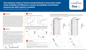
Poster: Gene expression analysis of immune checkpoint therapy in mouse tumor models reveals similarities and differences in immune cell populations and functional processes that reflect response to treatment
Presented at The Society for Immunotherapy of Cancer’s 35th Anniversary Annual Meeting & Pre-Conference Programs (SITC 2020)
in collaboration with Charles River
Experimental therapies that target the immune system have expanded greatly in recent years due to the success of immune checkpoint inhibitory antibodies such as ipilimumab and pembrolizumab. Preclinical development of these novel immune-oncology drugs requires the availability of well characterized mouse models to evaluate therapeutic mode of action, efficacy, and safety. Syngeneic mouse tumor models provide robust systems in which to evaluate novel immune-oncology therapies. Efficacy in these models can be measured by tumor volume changes in subcutaneous implants or by impacts on survival for orthotopic implants. Mode of action can be assessed by identifying changes in the tumor microenvironment following dosing. Multiple analytical methods can be used to track changes in immune populations and activation status from flow cytometry to immunohistochemistry to gene expression analysis. We endeavored to characterize the functional tumor microenvironment changes for two syngeneic models following treatment with anti-PD-1 and anti-CTLA-4 antibodies The syngeneic models used for the study were both colon adenocarcinomas, MC38 and CT26. Mice bearing subcutaneous tumors were dosed intraperitoneally with either vehicle alone, anti-CTLA-4, anti-PD-1, or a combination of the two immune checkpoint inhibitors on days 1, 4, and 8. Tumors were harvested on day 9 and assessed for gene expression by microarray analysis. The gene expression results were evaluated for the relationship between treatment regimen and tumor volume change by expression level association, functional set enrichment analysis, and immune cell population gene set variation analysis. For each of the tumor models, >10,000 genes were found to be significantly differentially expressed. Functional set enrichment analysis showed notable changes in cell cycle and mitotic markers as well as immune response markers in MC38. In contrast CT26 showed principally changes in immune response markers. Immune cell population set analysis revealed differential impacts on numerous immune cell populations between the models which correlate with therapy induced changes in tumor growth. These include expected changes in CD8+ T-cell populations for both models but also differential changes in other populations including CD56dim NK cells, eosinophils and B cells in MC38 and neutrophils in CT26. We also conducted a genomic analysis by whole exome sequencing. Both tumor models have relatively high tumor mutational burden; CT26 TMB = 377 MB and MC38 TMB = 69 /MB. These data show the value of robust bioinformatics analysis of gene expression data sets to provide insights into the mode of action and model responses to investigational immune-oncology drugs.


